

A Gallery: Neuroscientists in Color
Sarah McConnell, PhD
Neuroscientist
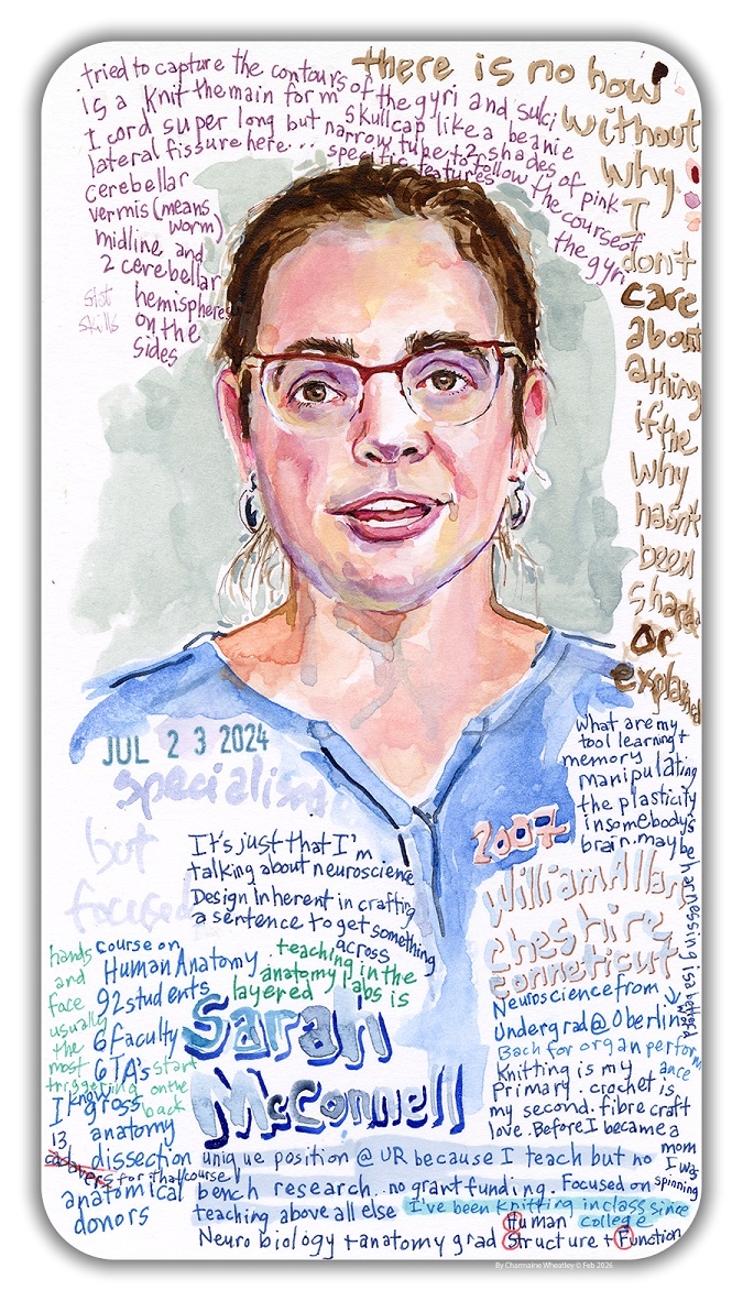
“Anatomy was not at all my plan in the beginning. I came to the University of Rochester for graduate school, thinking I would be an academic researcher. As a graduate student, I was advised to take that big medical school course, Human Structure and Function (HSF), so I did. I also completed my PhD, focusing on methamphetamine induced neurotoxicity, which was very much focused on the brain.
I remember being on cloud nine after being offered a full-time position at the University of Rochester. I was blindsided in the best possible way.
As I got to the end of my graduate school time, I realized I didn't love research. Things that didn’t really jive for me were the grant writing process and the fundamental injustice of the lack of alignment between your effort and your skill on the one hand and the outcome on the other. Research is trial and error. You may think something's going to work, and sometimes it just doesn’t. Some people don't get so frustrated by that, but I do.
I really wanted there to be alignment between how good I am at something, how hard I am working at it, and what I get out of it. I wanted something else, and I found that what really sparked joy for me was talking about science.
I knew then that I wanted to be a teacher, especially in higher education, where I could dive deep into science. The core of my teaching philosophy is neuroscience-based. As an educator, I’m trying to harness the synaptic plasticity in other people's brains.
After completing my PhD, I became an adjunct instructor in the area for a few years, which included teaching HSF at the School of Medicine and Dentistry.
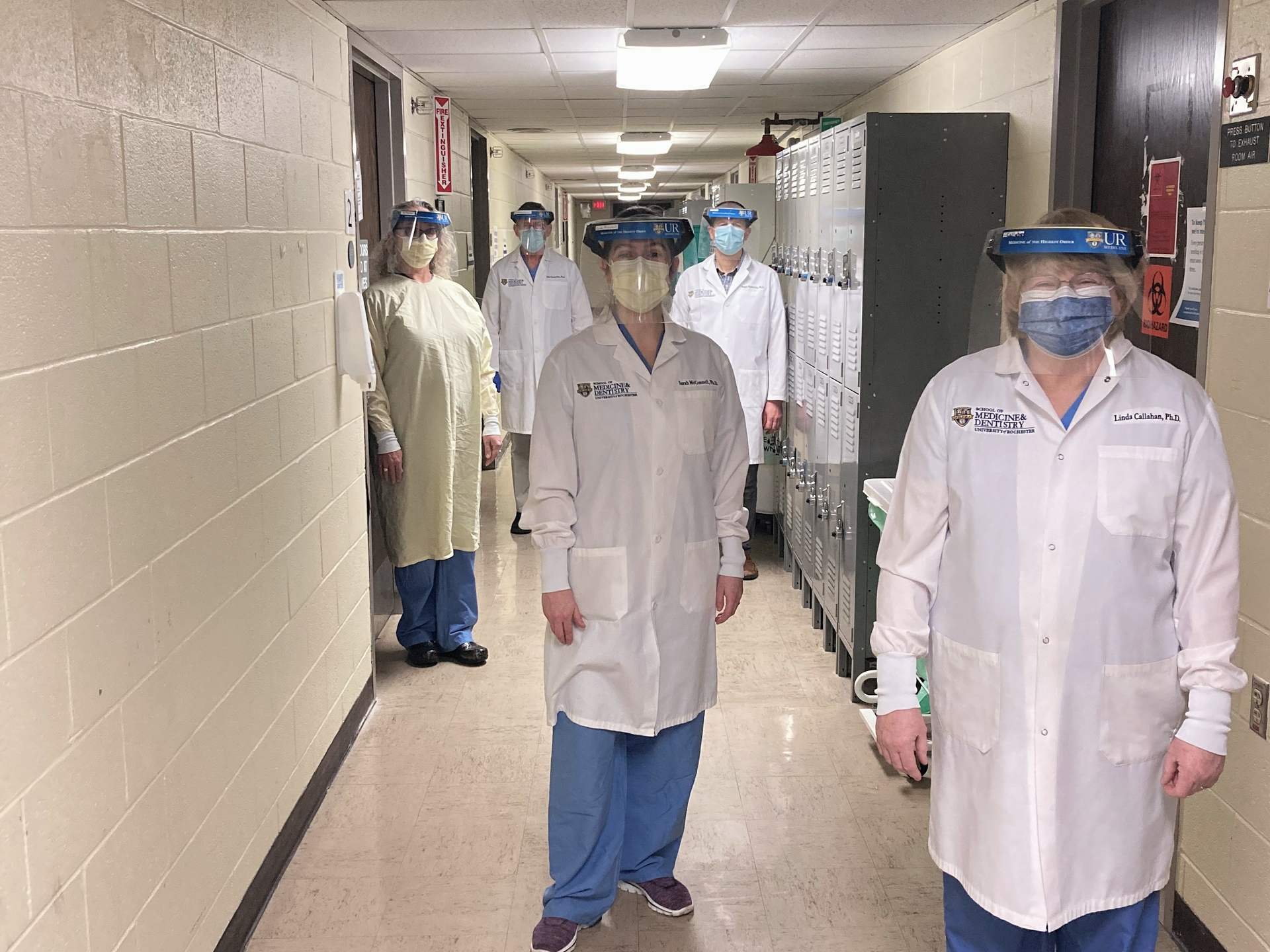
Suddenly, I was the director of this huge course, and things came full circle in a bunch of ways. I was teaching in the same place where I had learned. I was working with some of the same faculty who had taught me: Diane Piekut and Martha Gdowski, and now all of a sudden, we're colleagues and peers.
I go from talking to "Dr. Gdowski" to, like, "I guess I call you Martha now.”
It was completely wild getting myself back up to speed in time to teach this massive course with six TAs and eight faculty, and a real trial by fire. I guess it went well because they asked me to do it again the next year.
McConnell and Christopher Mooney published this unique hands-on activity lesson to teach embryonic lateral folding to medical students in Anatomical Sciences Education: https://doi.org/10.1002/ase.2062
I remember being on cloud nine after being offered a full-time position at the URochester. I was blindsided in the best possible way.
I had thought that to have a position at a place like the URochester, you had to be an R01 researcher. And I was like—that's not me. I don't want to do the grants. I don't want to do bench science. I want to teach.”
About the Artist
Charmaine Wheatley is the Artist-in-Residence (AiR) at the Del Monte Institute for Neuroscience at the University of Rochester, and holds a faculty appointment in the School of Medicine & Dentistry. She is a visual artist whose work is held in international museum and library collections, including New York’s Museum of Modern Art. She has received numerous awards and honors, and is a lifetime AiR at The Isabella Stewart Gardner Museum in Boston, MA. Her recent work at URMC has involved sitting with people from stigmatized communities - including those affected by Mental Health, HIV, and Opioid Use Disorder - and working to reduce stigma through her artistic practice of narrative portraiture.
Benjamin Suarez-Jimenez, PhD
Neuroscientist
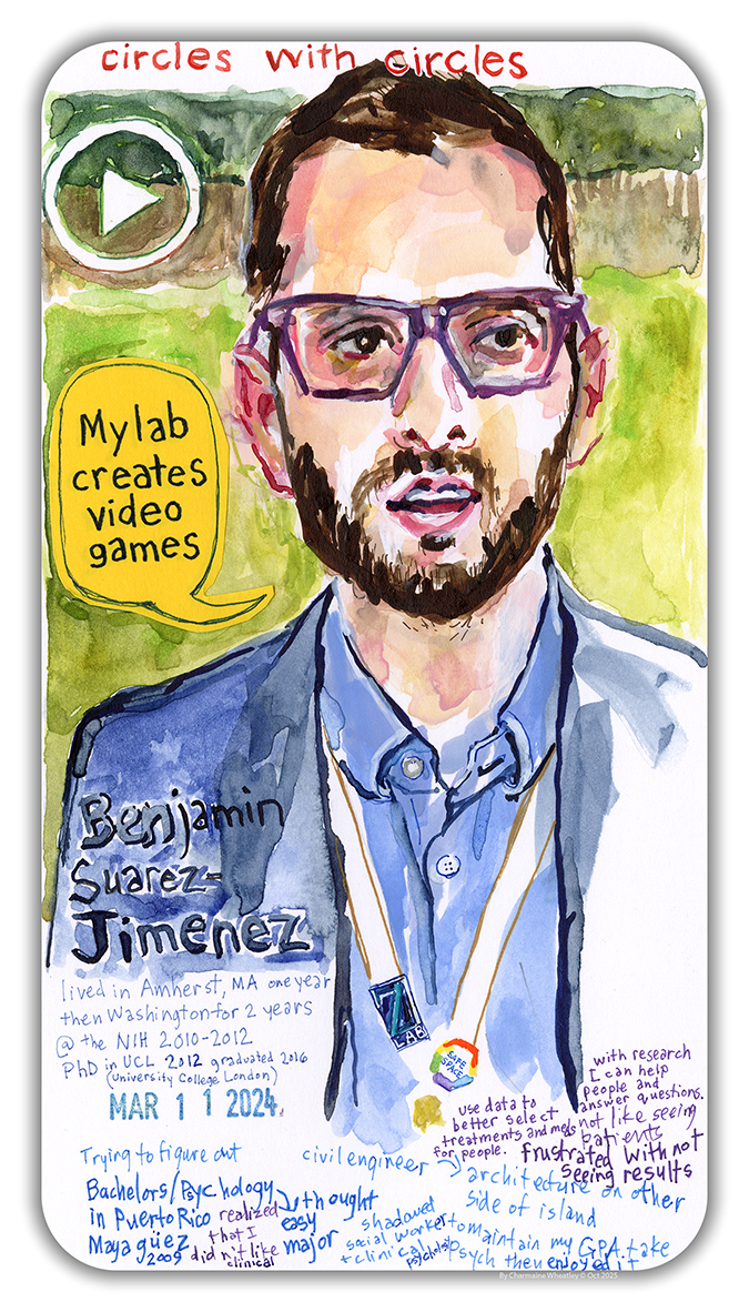
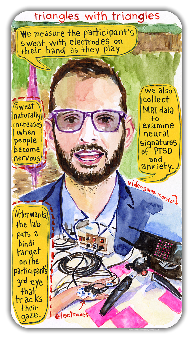
“I established the ZVR Lab at the University of Rochester to investigate the brain areas that build spatial maps and discriminate between the areas of an environment associated with emotions. Our lab uses virtual reality (VR) tasks, multimodal magnetic resonance imaging (MRI), eye-tracking, and machine learning methods to pinpoint the psychological and neural signatures of anxiety and trauma-related disorders like PTSD. Specifically, our goal is to understand how people build maps in the brain that are based on experience, and the role of those maps in psychopathologies of stress and anxiety. We are interested in cases where an area within a larger environment becomes associated with threat, such as the area of a neighborhood where an assault took place.
My primary objective is to illuminate the specific brain regions involved in discrimination learning and identify novel brain measures to contribute to the development of sensitive, personalized, and precise diagnostic and treatment tools for psychopathology. However, to better understand why PTSD develops and persists, we must first clarify how patients discriminate threat from safety and reward from non-reward within complex dynamic environments, by answering these critical questions:
- How do we form cognitive maps of threat and safety in our surrounding environment?
- How do these cognitive maps interact?
- How are these processes deficient in patients with post-traumatic stress disorder (PTSD)?
Virtual Reality Unlocks Clues
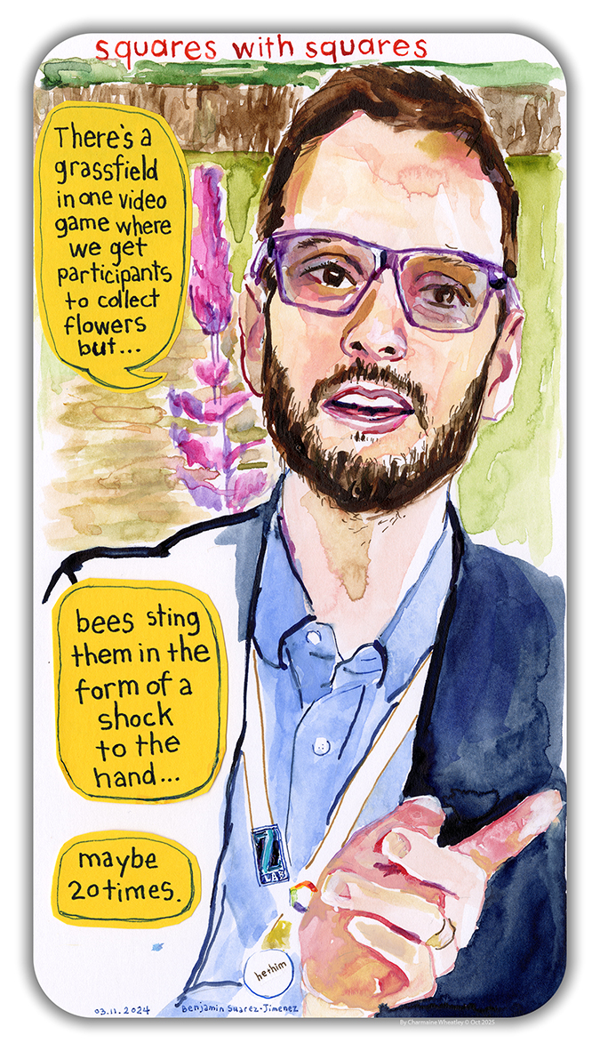
Virtual reality (VR) tools are ideal for assessing experiences in quasi-naturalistic environments. In the lab, we developed and use two novel VR tasks to assess brain activity differences between patients with PTSD, trauma-exposed controls without PTSD, and non-trauma-exposed healthy controls. In the Flower Task, a threat conditioning task, participants pick flowers in a virtual environment, and some of the flowers have a bee inside that will sting them. In the Moon Task, a reward-conditioning task, participants explore a virtual moon environment and collect fuel to return home.
Measuring attention
Another focus of my research is particularly directed toward the assessment of attention, learning, attention changes, and attention differences, utilizing advanced eye-tracking technology. Under the framework of my current R01 project, a free-viewing gaze tracking task is employed both before and after participants engage in the VR tasks that simulate threat and reward. We collect eye-tracking data as participants complete the virtual reality tasks inside the MRI.
Using the data gained from this research, we expect to contribute to the development of sensitive, personalized, and precise diagnostic and treatment tools for individuals who experience psychopathology.
Personal Experience Fuels Research
It was a combination of personal experiences, research I was doing as a post-doc, and my mentors. I’m from Puerto Rico and, as a teen, went to the only public boarding school at the time, which specialized in math and sciences. It was a stressful time for my peers and me. A lot of people dropped out, but it pushed me to do better. Looking back, it was the first time I started considering the types of questions about stress and anxiety that I’m asking today. Although I still didn’t go into science right away. I started my undergraduate studies as an engineering major before switching to psychology. That’s when I was introduced to research and began to get involved with projects related to resilience. During this time, my brothers enlisted in the military and were deployed to Iraq and Afghanistan. I saw how they were changing and eventually developed PTSD. I wanted to learn more about it and anxiety disorders in general, including how we can improve treatment and better diagnose these disorders.
These experiences as an early researcher solidified my commitment to understanding the neural mechanisms of environmental discrimination learning in individuals with anxiety and trauma-related disorders.”
About the Artist
Charmaine Wheatley is the Artist-in-Residence (AiR) at the Del Monte Institute for Neuroscience at the University of Rochester, and holds a faculty appointment in the School of Medicine & Dentistry. She is a visual artist whose work is held in international museum and library collections, including New York’s Museum of Modern Art. She has received numerous awards and honors, and is a lifetime AiR at The Isabella Stewart Gardner Museum in Boston, MA. Her recent work at URMC has involved sitting with people from stigmatized communities - including those affected by Mental Health, HIV, and Opioid Use Disorder - and working to reduce stigma through her artistic practice of narrative portraiture.
M. Kerry O'Banion, MD, PhD
Neuroscientist
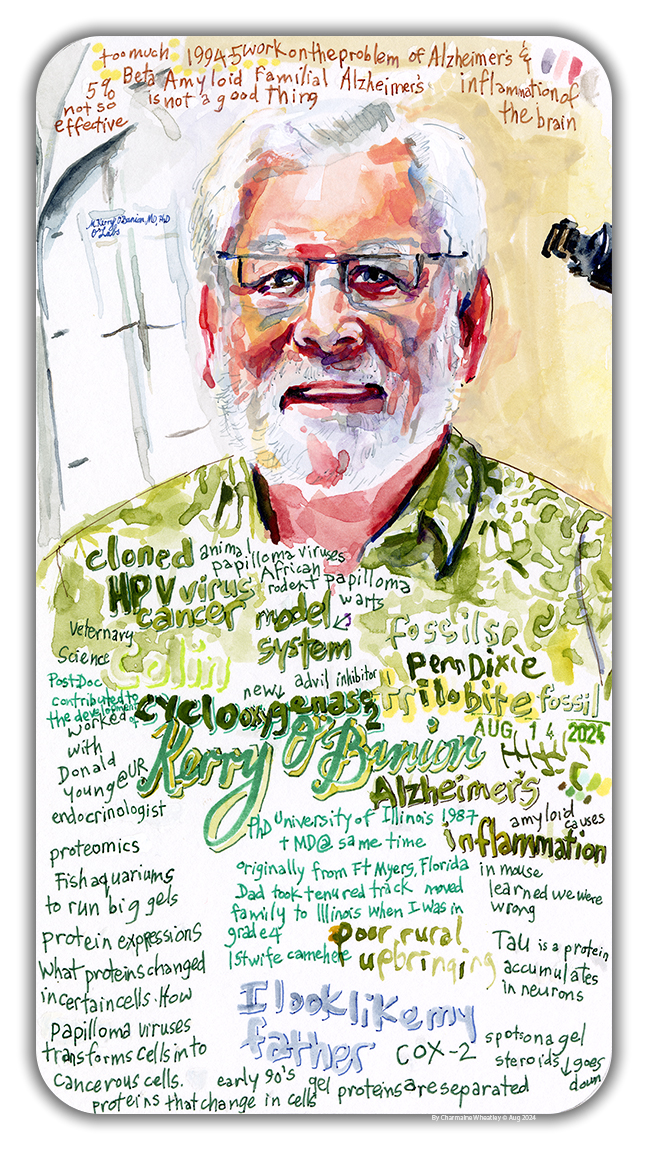
M. Kerry O'Banion, MD, PhD
Portrait by Charmaine Wheatley
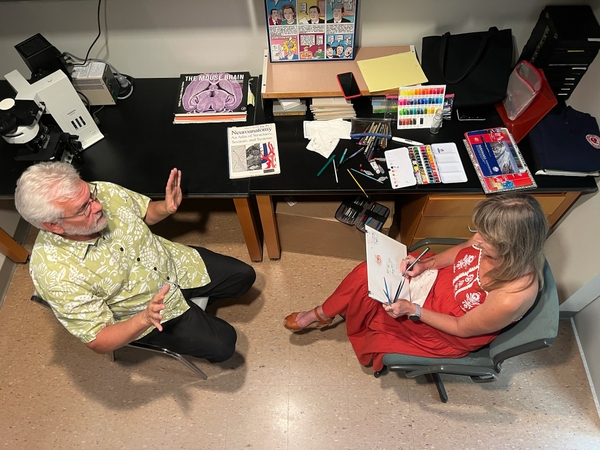
"As a newly minted MD-PhD trainee, I came to Rochester in 1987 to pursue research on wart viruses and their association with cancer. My postdoctoral fellowship was transformative and ended with me starting a faculty position in Neuroscience investigating new concepts about inflammation and Alzheimer’s disease. Nearly 40 years later, I’m still working on this problem: how does inflammation impact neurodegenerative disease, and what can we do about it?
We’ve learned that inflammation in the brain, or neuroinflammation, can be beneficial and detrimental. Moreover, we now know that cells and processes involved in neuroinflammation contribute to normal brain development as well as aging, and essentially all disease processes, whether instigated by injury, infection, toxins, or genes. Because of neuroinflammation’s widespread involvement in brain disease and normal function, my research program has expanded and led to multiple collaborations.
Alzheimer’s Disease (AD)
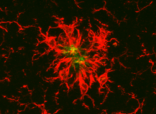
Nearly 25 years ago, we carried out a set of experiments that upended the then-current dogma that neuroinflammation drives AD pathology. Using mice developed to have localized chronic brain inflammation, we discovered that amyloid pathology was reduced and that this was due to microglia, the endogenous immune cells of the brain, phagocytizing or “eating” deposits of amyloid in the brain. In other experiments, we discovered that this same manipulation led to increased phosphorylation of tau, a pathological feature of AD and other dementing illnesses that causes neuron dysfunction and death. Thus, in trying to protect the brain from one pathology, microglial reactivity and accompanying neuroinflammation drives another pathology. With this in mind, together with our collaborator Ania Majewska, we have sought to identify the “good” neuroinflammatory factors, with the hope of enhancing these good actions without the bad. Currently, we are also testing whether microglia directly contribute to well-known sex differences in AD, with females showing greater disease burden than males.
Brain Radiation Injury
Rochester has a storied history of work in radiation biology, particularly in normal tissue injury. People who receive brain radiation as part of cancer treatment and survive often suffer from so-called “late radiation effects” that include loss of cognitive function. Together with colleagues in Radiation Oncology and Environmental Medicine, we have investigated the role of neuroinflammation in linking brain radiation injury to cognitive changes. Our work reveals a specific and potentially druggable function of microglia, namely the pruning of neuron connections, as a critical part of this link. Currently, we are working to identify radiation-driven biochemical pathways that cause these effects.
NASA and Space Radiation
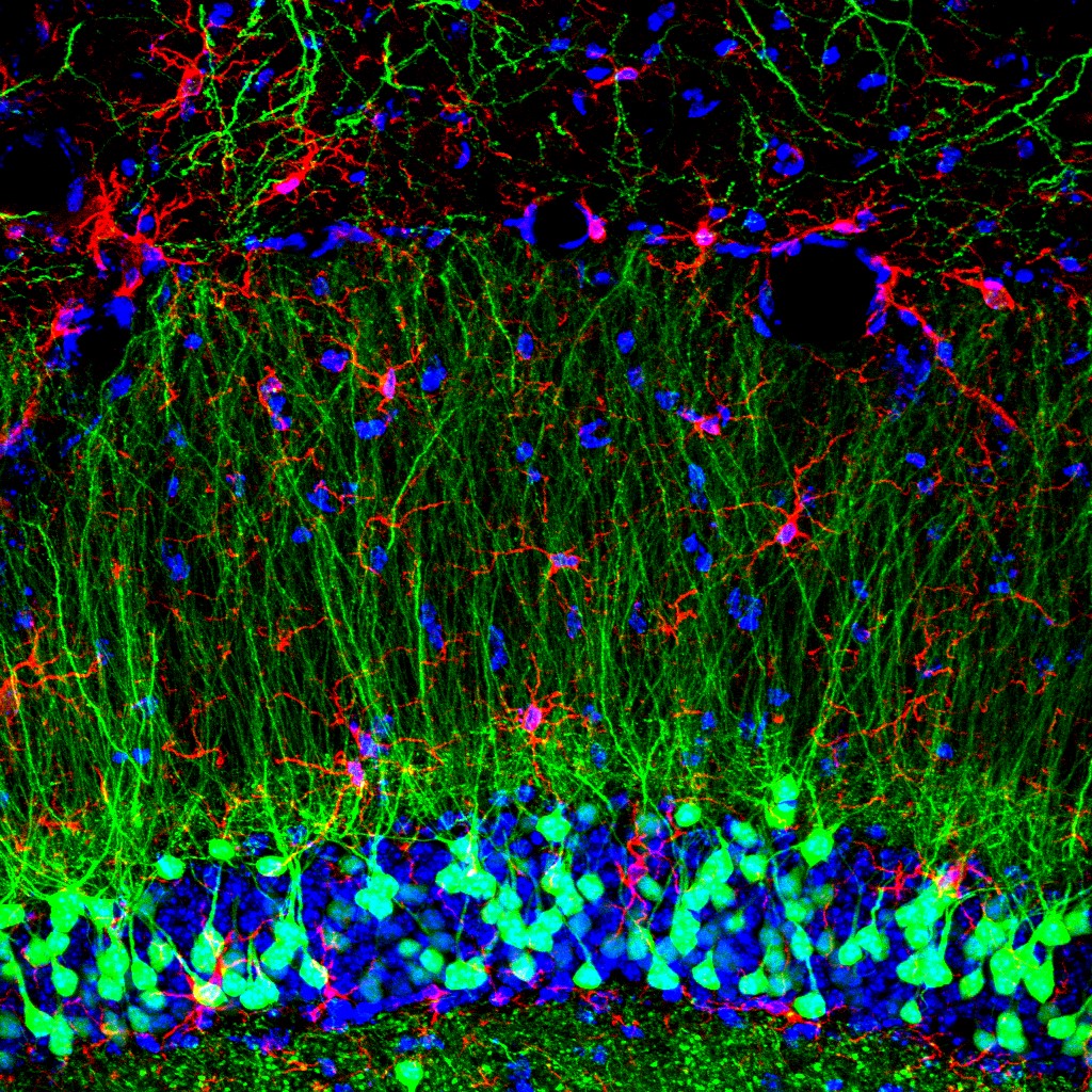
Our work on brain radiation injury in the context of cancer therapy has relevance for environmental radiation exposure, including challenges that will be faced by future astronauts traveling to the moon and Mars. Deep space, the area outside of low Earth orbit, which is protected from radiation by Earth’s magnetic field, is an environment with a high density of galactic cosmic rays, made up of small and large atoms moving at speeds approaching that of light, which arise from exploding stars. An additional risk is radiation from eruptions on the sun’s surface. With funding from NASA and access to a special facility at Brookhaven National Laboratory, we are studying the effects of “space radiation” on the brain, focusing on neuroinflammation, cognitive changes, and risk for neurodegenerative disease. Our current NASA project also includes a collaboration with Laura Calvi in the Department of Medicine and other members of the Wilmot Cancer Institute to investigate space radiation effects on the bone marrow and how this predisposes to blood cell cancers.
Aging Biology
Normal aging is associated with increased inflammatory burden, including in the brain. We recently started collaborations with Vera Gorbunova and Andrei Seluanov, professors in the Department of Biology, who use comparative biology to understand why some animal species live longer than others. Our goal is to learn whether genes involved in increasing life and health span might also impact the progression of neurodegenerative diseases. We explore this goal using genetically modified mice and a unique rodent species, the Degu, that models sporadic, late-onset AD."
About the Artist
Charmaine Wheatley is the Artist-in-Residence (AiR) at the Del Monte Institute for Neuroscience at the University of Rochester, and holds a faculty appointment in the School of Medicine & Dentistry. She is a visual artist whose work is held in international museum and library collections, including New York’s Museum of Modern Art. She has received numerous awards and honors, and is a lifetime AiR at The Isabella Stewart Gardner Museum in Boston, MA. Her recent work at URMC has involved sitting with people from stigmatized communities - including those affected by Mental Health, HIV, and Opioid Use Disorder - and working to reduce stigma through her artistic practice of narrative portraiture.
Ania Majewska, PhD
Neuroscientist
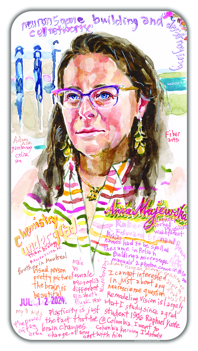
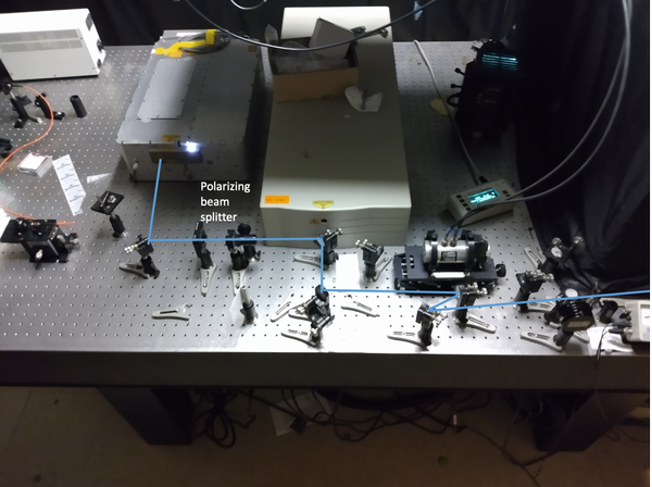
"I went to Columbia University in 1996 as a graduate student knowing I was going to study with Rafael Yuste. That's why I went there. It was because of the new science that was enabled by two photon microscopy.
Dr. Yuste studied how the connections between neurons change, which is the remodeling and the plasticity that really drives all of my work. He was studying it in the visual system - and a lot of the insights that are learned from the visual system can be applied broadly to the brain.
Microglia
Microglia are immune cells. They're not even brain cells. They’re actually born outside of the fetus, in the yolk sac. And then they come in very early and they colonize the brain, and become these resident immune cells of the brain.
👀 Watch microglia move in the brain 👇
Even though they're immune cells - cells that are supposed to be involved in injury, disease and inflammation - it seems that they have normal roles in the healthy brain.
In fact, they are constantly moving. They're beautiful. They’re dynamic. And they're constantly touching these points of connections between neurons. What’s happening when they do that, is that they are helping the brain learn and constantly remodel these neural networks.
Brain Plasticity and Remodeling
It blows my mind that I can look at these deep brain structures and connections that are smaller than a micron. I can see a structure, and two hours later that structure’s gone. The whole connection is gone. Those two neurons are no longer talking to one another.
👀 Watch as microglia react to an injury 👇
Those connections have really impressive, molecular infrastructure in them. They're complicated. It's not easy to build one, but the brain is constantly building and destroying them as a way to change the way the neurons talk to one another, and to build, modify and rebuild neural networks.
Why This Matters
Changes in the way neurons communicate are crucial to brain function, including development, learning and aging. Defects in this neuronal plasticity underlie many neurological disorders, such as autism, epilepsy and Alzheimer’s disease.”
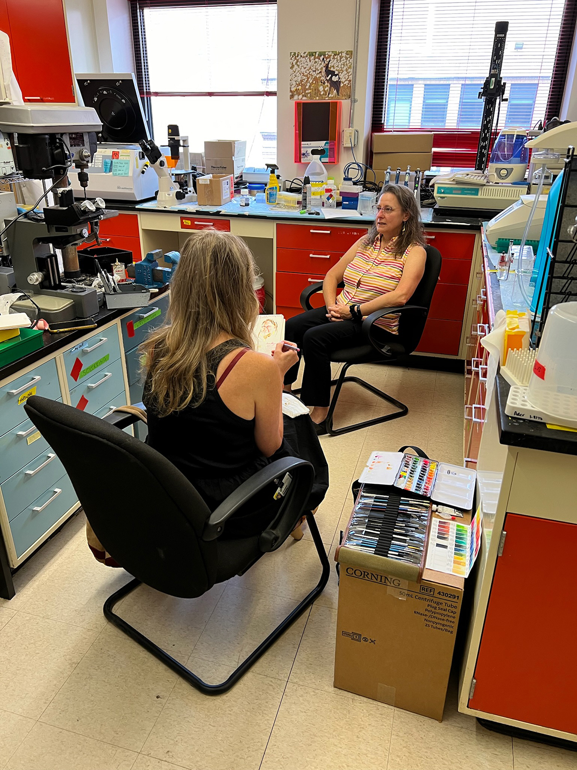
Learn more: Majewska Lab
About the Artist
Charmaine Wheatley is the Artist-in-Residence (AiR) at the Del Monte Institute for Neuroscience at the University of Rochester, and holds a faculty appointment in the School of Medicine & Dentistry. She is a visual artist whose work is held in international museum and library collections, including New York’s Museum of Modern Art. She has received numerous awards and honors, and is a lifetime AiR at The Isabella Stewart Gardner Museum in Boston, MA. Her recent work at URMC has involved sitting with people from stigmatized communities - including those affected by Mental Health, HIV, and Opioid Use Disorder - and working to reduce stigma through her artistic practice of narrative portraiture.
Nathan Anthony Smith, PhD Neuroscientist
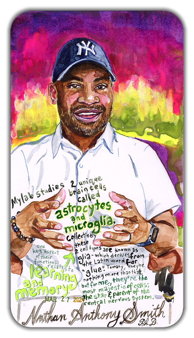
"My lab studies two very unique brain cells called astrocytes and microglia. Collectively, these two cell types are known as glia – which derives from the Latin word for “glue”. To many, they’re nothing more than that, but to me, they are the most majestic of cells; the star and parent of the central nervous system. These cells are critically important to how our brains work – both in health and in disease.
One key aspect of their function relates to learning and memory. Many scientists think of these defining aspects of sentient life as being mediated by neurons (aka the principal “thinking” cells of the brain). And it’s certainly true that learning and memory require the formation of new neural networks however, glial cells play a critical role in how those networks are formed, nurtured and maintained.
The complex branching and interweaving of neural networks, and their ability to adapt and remodel is a core feature of neural plasticity – a concept that underlies our ability to think, remember and learn. But neural plasticity is not solely a function of neurons.
Changes in neural connections, and the remodeling of the dendritic arbor, require the action of both microglia and astrocytes. Microglia are the resident gardeners of the brain. They prune the branches of the tree – removing synapses (neural connections) that are no longer needed and permitting the addition of new synapses. Astrocytes help strengthen synaptic connections that are necessary for new neural pathways to form – and for learning and memory to develop. Collectively, astrocytes, microglia and neurons act collaboratively to make us fully human.
My scientific interests include elucidating how astrocytes and microglia play a role in multiple processes, including information processing, and synaptic plasticity – and in learning and memory. An additional goal of my research is to discover and understand new pathways that we can target therapeutically. We can start to look at glial cells as therapeutic targets, rather than looking only at neurons. To do that, we need to figure out how the whole system works – which is what my research focuses on- with the long-term aim of finding new therapeutic targets that can solve neurological diseases, and improve the quality of life of affected people."
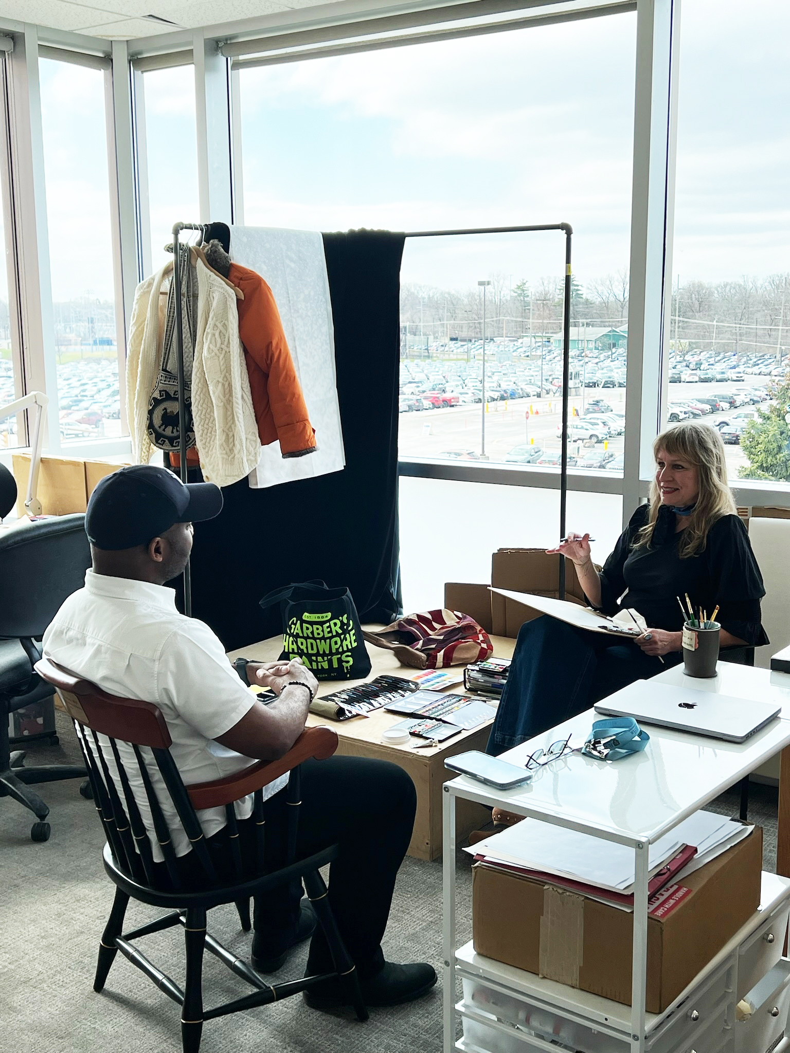
Nathan A. Smith, MS, PhD ('13), sits for his portrait with Charmaine Wheatley, artist-in-residence at the Del Monte Institute for Neuroscience.
Learn more: The Smith Lab
About the Artist
Charmaine Wheatley is the Artist-in-Residence (AiR) at the Del Monte Institute for Neuroscience at the University of Rochester, and holds a faculty appointment in the School of Medicine & Dentistry. She is a visual artist whose work is held in international museum and library collections, including New York’s Museum of Modern Art. She has received numerous awards and honors, and is a lifetime AiR at The Isabella Stewart Gardner Museum in Boston, MA. Her recent work at URMC has involved sitting with people from stigmatized communities - including those affected by Mental Health, HIV, and Opioid Use Disorder - and working to reduce stigma through her artistic practice of narrative portraiture.
